

获取更好蛋白质结构的新方法
来源:《Science》
作者:Wei Liu等
时间:2014-03-12
确定大蛋白三维结构的改良方法似乎比传统技术更能展现蛋白结构的自然姿势。
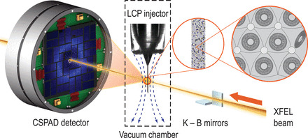
称为G蛋白偶联受体(GPCRs)的蛋白质是重要的药物靶标,但研究人员苦于弄清楚其结构。加利福尼亚斯克里普斯研究所Vadim Cherezov和同事利用X射线自由电子激光器改良了标准的X射线晶体学技术,捕获神经递质羟色胺的G蛋白偶联受体的系列结构图像。
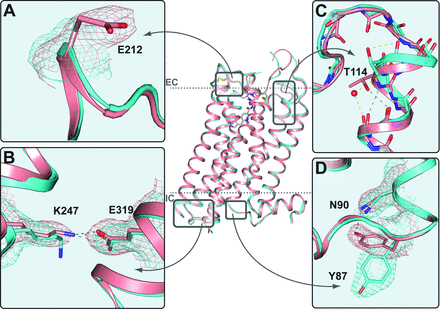
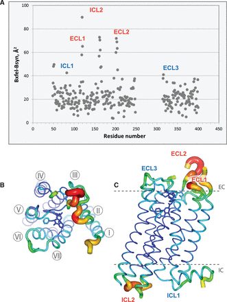
在室温下保存的微小5-羟色胺受体晶体上,研究团队使用该技术得到的结构不同于那些使用常规方法探测获得的冷却较大晶体结构。结果表明,室温下自由电子激光方法可以更好地捕捉自然环境中的蛋白质构象。(编译:中国科学院成都生物研究所 王芋华,王海燕)
Serial Femtosecond Crystallography of G Protein–Coupled Receptors
Abstract X-ray crystallography of G protein–coupled receptors and other membrane proteins is hampered by difficulties associated with growing sufficiently large crystals that withstand radiation damage and yield high-resolution data at synchrotron sources. We used an x-ray free-electron laser (XFEL) with individual 50-femtosecond-duration x-ray pulses to minimize radiation damage and obtained a high-resolution room-temperature structure of a human serotonin receptor using sub-10-micrometer microcrystals grown in a membrane mimetic matrix known as lipidic cubic phase. Compared with the structure solved by using traditional microcrystallography from cryo-cooled crystals of about two orders of magnitude larger volume, the room-temperature XFEL structure displays a distinct distribution of thermal motions and conformations of residues that likely more accurately represent the receptor structure and dynamics in a cellular environment.
原文链接:http://www.sciencemag.org/content/342/6165/1521.full.pdf